BioAcyl Corp |
|
Tan, J., Anderson, D. E., Rathore, A. P. S., & others. (2021). Signatures of mast cell activation are associated with severe covid-19. medRxiv, 2021.05.31.21255594. Added by: Dr. Enrique Feoli (05/06/2021, 08:10) Last edited by: Dr. Enrique Feoli (05/06/2021, 10:15) |
| Resource type: Journal Article BibTeX citation key: Tan2021 View all bibliographic details |
Categories: BioAcyl Corp Subcategories: COVID-19 Creators: Anderson, others, Rathore, Tan Collection: medRxiv |
Views: 4/218
|
| Abstract |
|
Lung inflammation is a hallmark of Coronavirus disease 2019 (COVID-19) in severely ill patients and the pathophysiology of disease is thought to be immune-mediated. Mast cells (MCs) are polyfunctional immune cells present in the airways, where they respond to certain viruses and allergens, often promoting inflammation. We observed widespread degranulation of MCs during acute and unresolved airway inflammation in SARS-CoV-2-infected mice and non-human primates. In humans, transcriptional changes in patients requiring oxygen supplementation also implicated cells with a MC phenotype. MC activation in humans was confirmed, through detection of the MC-specific protease, chymase, levels of which were significantly correlated with disease severity. These results support the association of MC activation with severe COVID-19, suggesting potential strategies for intervention.
|
| Notes |
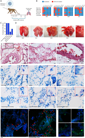
Widespread activation of MCs coinciding with lung pathology in NHPs. (A) Cynomolgus macaques were infected intratracheally with SARS-CoV-2 and monitored for 21 days prior to necropsy. (B) Viral detection was determined by PCR at regular intervals post-infection in swabs from multiple mucosal tissues, lung lavage, and nasal rinses. All NHPs were positive for SARS-CoV-2 infection multiple days after inoculation. (C) Abnormal findings related to lung tissue observed at the time of necropsy were recorded and effected all animals. (D) Images of NHP lungs at the time of necropsy show areas of hemorrhaging and necrotic spots on the lung surface. Boxed region is enlarged. (E) Histological assessment of lung tissues by H&E staining shows hemorrhaging of the tissue and free RBCs within the lung alveolar spaces. (F) Inset corresponding to the boxed region of panel H. (G) Some RBCs in the tissue proximal to a blood vessel are indicated by arrows and cellular infiltrates are circled. (H) Multiple examples of degranulating or hypogranulated MCs are provided, observed in toluidine blue stained lung tissue sections. The MCs are enlarged in the red-outlined insets. For (I-K), lung sections were stained for MC heparin to indicate the location of MC granules (green) and DAPI to identify cellular nuclei and tissue structures. MCs are indicated with red arrows. (I) MCs were observed degranulating in the lung of SARS-CoV-2 infected primates in sections of a biopsy of lung tissue that did not have overt hemorrhaging visible on the lung surface at necropsy. (J) MCs appear more densely packed in the lung biopsy from a hemorrhagic lobe of the lung and again, degranulation is observed based on staining for MC-heparin. (K) Images of degranulating MCs are presented at higher magnification.
Added by: Dr. Enrique Feoli Last edited by: Dr. Enrique Feoli |